Phone: +7 (383) 330-67-71, Fax: +7 (383) 330-80-56, E-mail: bic@catalysis.ru
5 Lavrentiev Ave., 630090, Novosibirsk, Russia
Phone: +7 (383) 330-67-71, Fax: +7 (383) 330-80-56, E-mail: bic@catalysis.ru
5 Lavrentiev Ave., 630090, Novosibirsk, Russia
Phase Analysis Methods
Single- and multi-component polycrystalline and nanocristalline systems.
X-ray powder diffraction is used for the study of the composition, structure, microstructure, and phase conversion dynamics in the single- and multi-component polycrystalline and nanocristalline systems.
Following estimations are performed:
dynamics of phase composition variation, dispersity, structural parameters of specific phases (unit cell parameters, structural type, characteristics of an atomic structure deviation from the ideal one under the temperature and medium influence and others), kinetic parameters of phase transitions.
The method becomes a necessity under the reversible action of the specific conditions onto the substance and might be used in the various fields of science and engineering (catalysts, minerals, metals and any chemical compounds).
1. In the Siberian Center of Synchrotron Radiation of the Siberian Branch of the Russian Academy of Sciences (SB RAS) jointly with the Institute of Chemistry of Solid and Mechanochemistry of SB RAS a precise diffractometer allowing to obtain the diffraction patterns with a resolution (peak half width) up to 0.03° by 2Θ in the wide range of wave length 0.05 – 0.4 nm. Due to the possibility of wave length variation the anomalous scattering method might be used for the study of isomorphous substitutions and for the construction of the partial functions of radial atom distribution.
2. An original software for the simulation of real structure and diffraction patterns of ultra-dispersed and partially disordered systems has been developed. With this technique on the basis of a full profile analysis of the diffraction pattern it is possible to obtain the detail information about CSD, micro-strains (I and II type), type and concentration of defects of layers deposition and others. It is possible to study the real structure of a lot of objects, including crystals with a developed micro-domain structure and crystals with very small physical sizes, paracrystals, turbostrate structures, various polytypic and modulated structures as well as diversity of real systems in which various sources of disordering of the long-range order simultaneously take place.
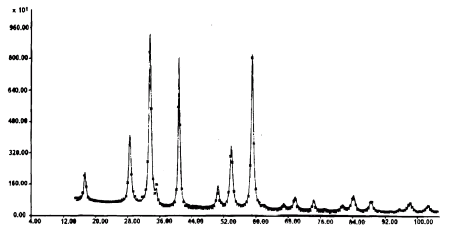
Experimental (asterics) and theoretical (solid line) X-ray diffraction patterns
of aluminum-magnesium spinel (R1 = 4.5 %, Rp = 7.6 %)
A full profile analysis of X-ray diffraction images allows not only to refine the average crystalline structure (atoms coordinates in the elementary cell, positions occupation, heating parameters) but to define the structure of extended defects as well. The example given for the non-stoichiometric aluminum-magnesium spinel displays these possibilities. The theoretical diffraction image corresponds to the experimental X-ray pattern with a high accuracy.
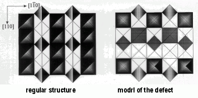
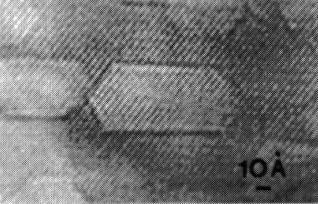
The local structure and concentration of the planar defects are determined from the X-ray diffraction data. They constitute the dislocation walls forming the pseudo hexagonal closed loops that are visualized by the technique of high resolution electron microscopy (HRTEM).
/Journ. Struct. Chem. [JSTCAM], v.32, p.325 [1997]/
Siemens D500, URD-6, URD-63, HZG-4B, HZG-4C, Bruker D8 diffractometers (Germany); high resolution diffractometer with synchotron emission in the Siberian Center of Synchotron Radiation of SB RAS. The X-ray tubes with the copper and molybdenum anodes are applied on the series-produced diffractometers. Bases of X-ray diffraction powdery (PC-PDF) and crystallostructural data are available.
Samples in the form of powder (0.5–2 cm3), plates, tablets (not less than 5x5 mm2) with the indication of chemical composition.
Prof. S.V. Tsybulya. Full profile analysis and study of the defective structures.
Prof. L.M. Plyasova. Regularities of the formation of complex oxide catalytic systems, X-ray in situ.
Prof. E.M. Moroz. Study (including by REDD method) of the structure of oxides, carbon-containing materials, highly dispersed bulk and supported metals.
Solid and fluid samples in the form of surface slicks, plates, coatings, powders, tablets, pellets, solutions, sols and gels.
Small-angle X-ray diffractometer of Siemens (Germany), Anton Paar and Hecus-Braun firms (Austria) with small-angle camera Kratky (“Compact camera”). The camera allows to measure X-ray scattering on solid and liquid samples in the area from the smallest angles up to 7 grad; the maximum resolution of the camera is 300 nm; the X-ray tube with a copper anode (λCuKα = 0.154 nm) is applied; there is ability of samples termostabilization from 0 up to 70°C within ± 0,1°C.
Thin films, tablet, plate, coating by the size not less than 1x10 mm2; powders, pellets, solutions, gels and sols with a volume of a disperse phase not less than 0.05 cm3.
Prof. F.V. Tuzikov. Study of molecular mechanisms of chemical and biological catalysis, nanostructural analysis of substances, analysis of the components of blood for medicine, small-angle radiography in situ, development of scientific equipments.
The method may be used in regulation of phase composition during preparation and exploitation of the heterogeneous catalysts, high temperature superconductors, luminescent solids, magnetic materials, inorganic pigments, products of mechanochemical activation, for the study of archeological findings and others.
Simultaneous phase classification of 38 chemical components is possible.
The sensibility of component determination is up to 10–3 mkg/ml.
An error of the stoichiometric coefficient estimation in the phase formula is up to 10%.
Method uniqueness: determination of the empirical phase formula and their quantitative content in multi-phase samples is performed without reference samples of the corresponding individual phases.
The method is based on the regularities of stoichiography and dynamic diffusion regime. Inductively coupled plasma atomic-emission spectrometer (PST “Baird”) is used as a detector.
Sample weight – from milligrams up to grams.
Prof. V.V. Malakhov and Dr. A.A. Vlasov. Phase analysis of the solid inorganic substances.
X-ray amorphous liquid and solid samples, i.e. the samples for which X-ray diffraction structural methods are unsuitable.
The structure of the local atom surroundings of the selected chemical component (coordination number, interatomic distance, Debye factor, type of the neighboring atom) is studied. Depending on the technique applied the following parameters are analysed: volume, surface or near the surface layers.
| Chemical components to be studied | |
| Concentration of the component to be studied, % mass | |
| Range of the interatomic distances to be studied, nm | |
| Error of the coordination number determination, % | |
| Error of the Debye factor determination, % |
Institute of Catalysis - a manager of the unique EXAFS spectroscopy station in Russia – specializes in the study of highly dispersed objects – catalysts, nano-materials. The procedures of sample preparation are developed for surveying the reaction-active compounds and catalysts under the inert conditions. The sample study is possible at the temperature from 77 to 900 K under the preset atmosphere conditions.
The station is mounted at the VEPP-3 storage ring located at the Institute of Nuclear Physics of the Siberian Branch of the Russian Academy of Sciences with an electron energy of 2 GeV. The following techniques have been realized: “on transmission”, X-ray fluorescence, total reflection, total photocurrent, X-ray stimulated optical luminescence.
Sample – solid or liquid – in a quantity of 20 mg with respect to the chemical component to be studied. The solid samples – in the form of powder particle size not more than 0.1 mm. Regarding all the samples the preliminary consultation is necessary. In case of the sample study in inert conditions the ampoules are presented or the consultation about their preparation is provided.
Prof. D.I. Kochubey and Dr. V.V. Kriventsov. Study of the ultra-dispersed metals, cluster, oxide and sulfide materials.
Prof. D.I. Kochubey is the author of monographs “EXAFS spectroscopy” (1988) and “EXAFS catalyst spectroscopy” (1992), in Russia.